Labels

Wednesday, 2 October 2013

BYE BYE painful injections for dental treatment...! 


Hello friends, today i would like to offer everyone an insight on the comparable and needle free pain control methods in dentistry.

1) Electronically delivered anesthesia (also called trans-cutaneous electrical nerve stimulation [TENS]).

This is an alternative to the shot of anesthesia.
Adhesive pads are placed on the face and a battery-powered device sends electrical impulses to the treatment area to numb it. The patient and the doctor can control the level of stimulation through a hand-held unit.
Another form of electronically delivered anesthesia is called cranial electrotherapy stimulation. Under this technique, electricity is passed into the brain, which causes relaxation.
Again, the patient or doctor controls the intensity of the current, increasing or decreasing it to control the pain as needed.

Advantages:- as soon as the device is switched off, the effect of anesthesia diminishes almost instantly unlike LA.  And the patient is able to drive and resume normal activities immediately following the dental visit unlike sedation or GA.


2)LASER drills:- Some dentists are now using lasers to remove decay within a tooth and prepare the surrounding enamel for receipt of the filling. cavity prep. with LASER is painless and thus no need for anesthesia .

=> other methods like performing procedure under:--
3) N2o (laughing gas)
4)oral conscious sedation
are also effective in pain control but have their own merits and demerits.

also some methods which are not yet clinically proven have been advised like => acupressure and acupuncture  hypnosis etc

so as science advances , newer methods of pain control may finally help to remove needle phobia amongst patients and make them seek dental treatments promptly :)

Sunday, 19 August 2012


Donate your child's teeth and save lives


The next time your child's milk tooth is about to fall, instead of summoning the tooth fairy, donate it to a stem cell bank -- which could help treat a range of diseases including parkinsons and alzheimers in adults.

Stemade has opened their first dental stem cell bank in the city where it is now possible to freeze your dental stem cells for future use. The bank, which is functioning in Mumbai, Delhi, Pune, Bangalore and Hyderabad, has now opened a facility in Chennai, too.

Dental surgeon and oral pathologist, Dr K Ranganathan said that adult stem cells are of two types, hematopoietic stem cells that form blood cells and mesenchymal stem cells that form connective tissues. "Dental stem cells are mesenchymal stem cells that can be potential treatment options in diseases like parkinsons and alzhemiers," he said. He also added that dental stem cells, because of their place of origin, were more versatile than hematopoetic cells. "The jury is out on this, but we feel dental stem cells are more competent," he said.


Dr Ranganathan also said that the biggest advantage of the dental stem cell banking was that it was a non-invasive procedure and the teeth would be of no use to the person who would anyway throw them away. "Besides it is a great opportunity for people who have missed out on banking their cord blood cells to bank their dental stem cells, as dental stem cells can be extracted at any stage," he said.

Managing director of Stemade Shailesh Gadre said that unlike umbilical cord cells which are used to treat blood-related diseases like leukaemia and thalessemia, dental stem cells have the ability to treat certain forms of cancer, heart disease, periodontal diseases and can also be used to grow replacement teeth and bone. He added that the procedure of extraction would have to be done while the milk teeth was about to fall or when the wisdom teeth were about to be extracted. "The dental pulp of the teeth is the source of these stem cells and the pulp is more usable when they have a blood supply," he said. He also added that dental stem cells have high regeneration capacity and multiply fast.
Stemade has partnered with seven dental clinics in the city which are equipped with all the facilities to perform dental stem cell extraction. The cost, said Gadre, would work out to Rs 1.5 lakhs for the 15-year period for which the dental stem cells were being stored. "The processing fee is Rs 60,000 and the banking cost is Rs 6,000 per year," he said.

Whole teeth grown from molar stem cells in mice

     Few months ago in my blog , i had discussed about how close researchers are to develop technique of growing teeth by genetic engineering. Friends that time has come really fast ......

actual photgraph of implanteed mandibular teeth


Researchers in Japan have grown functional teeth using cells taken from a mouse molar as starting material. The group grew these cells for a few days in the lab, then placed them into a tooth-shaped mold and implanted the mold into the mouse kidney where they were left to grow for about two months.

What the group found after that time looked like a tooth, had the normal structures of a tooth and was able to implant normally into the jaw of a mouse. The mouse was able to chew normally using the engineered tooth.

The work was published on July 12 in the journal PLoS ONE. In a Reuters story about the work,Takashi Tsuji, who led the research, said two things are needed before dentists can replace artificial bridges with teeth that implant and function just like the original set. One is learning how to grow the teeth in a lab rather than inside the kidney.

The other is finding the human equivalent of the cells they took from mouse molars. Molars contain a lot of cells, only some of which have the ability to create entire new teeth under the right conditions. In the Reuters story Tsuji said:
"In this case, entire tooth units could be grown because the stem cells were taken from molar teeth of mice -- where they later grew into enamel, dental bones and other parts that comprised a regular tooth unit."
As is often the case in medical research, translating discoveries from mice or rats to humans can be slow going. However, if the scientists are able to find the right cells and work out conditions to grow those cells in the lab, in could mean a future of easier chewing for older people.

Friday, 17 August 2012

DENTAL PHOBIA


It is a fact that many people are still frightened to go to the dentist but dental phobia is the main cause of deteriorating dental health.

Causes of dental phobia:

Uncomfortable or traumatic past dental experience:
The most common cause of dental phobia; and it is thought to be the most difficult to overcome if it happened in early childhood and contributes to a deep-seated fear of dental intervention.
Lack of communication:
Anxious dental patients need continuous explanation and reassurance. If adequate time is not taken to explain, respond to concerns and answer questions; patients often become disillusioned, confused and unsure about the nature and outcome of treatment. Consequently, anxiety and fear increase to the extent that the patient would not return to the dentist, or visit any other dentist!
Invasion of personal space:
The mouth is a body cavity; it is an intimate personal space, therefore, some individuals feel extremely anxious about a very close “invasion” of such a personal space.
Fear of needles:
Also known as needle phobia. Individuals with needle phobia will not seek dental treatment, as they are frightened of the dental injection (or any form of injection).
“I let my teeth deteriorate, I am ashamed of them, I think I am going to have false teeth”:
Some patients who neglected their dental health for reasons other than fear (demanding work, caring for children, elderly or disabled family member..Etc.) believe that the dentist would be astonished and would remove all their remaining teeth and replace them with dentures. These individuals have a morbid fear of losing their own teeth and having dentures.
Negative or exaggerated remarks from friends or relatives who may not have had a bad experience themselves!
5 TOP TIPS TO OVERCOME THE FEAR OF DENTISTS

First, you have to realise that there are thousands of individuals like yourself; dental phobia is like any other fear; for example fear of heights or closed spaces. You are not on your own.
Second, you also have to realise that there are professionals who can help you overcome your fear. Through good communication, explanation and reassurance, your fears will tend to reduce, and you will be more in control of your anxiety.
Third, dental anxiety management techniques are now well developed and highly successful. Dental practitioners who practice anxiety management would help you overcome your fears through the administration of certain medications.
Forth, Find an understanding and sympathetic dental practitioner. Usually you would sense the mannerism in the practice from the front desk. If the atmosphere is welcoming, relaxed and courteous, and the staff provide you with enough information and respond to your enquiries; the chances are that you are in the right place.
Fifth, let the staff and dentist know of your dental anxiety, and enquire if they practice anxiety management techniques.
Finally, I  MYSELF FEARED DENTAL TREATMENTS INSPITE BEING A DENTAL STUDENT BUT  I have to assure all of those who are reading this article and suffer from dental phobia, that help is available, it just takes this first step; making contact, and you may be surprised how confident and comfortable you will feel afterwards :)))

Sunday, 8 January 2012

Surgical Repair of Post Perforation


Surgical Repair of Post Perforation

This patient presented without pain but presence of draining sinus tract in the buccal attached tissue over #7. The position of the draining sinus tract directly at the level of the post, normal periodontal probings and evaluation of the radiograph made me suspicious of a post perforation. (note the widened ligament to the level of the post, and completely normal appearance at the periapex)
Options discussed included retreatment or apical surgery. Since the crown is all porcelain, we decided to treat surgically.

Ochsenbein-Luebke surgical flap was selected to prevent recession of marginal gingival, and minimal loss of crestal bone. The periodontal attachment was still in tact following flap reflection despite the loss of buccal bone adjacent to the perforation.


Metal tip of the post was visible without any removal of any buccal bone.

The post was counter-sunk using a high speed handpiece.

Preparation of the root. Note that this was not the apex of the root. This repair was being done on the mid-root surface.


Geristore was selected as the restorative material. Since Geristore is a bonded material, moisture control is important. Astringedent was used for hemostasis and the root was acid etched, primed and bonded.

Geristore placed in the preparation and cured.

Geristore was contoured to the root surface.


Final film. This will be an interesting case to follow. Expect a good result and repair of boney defect. We'll continue to monitor this tooth over time. Endodontic surgery has provided a valuable service to this patient prolonging the life of this tooth, the crown and hopefully regenerating the boney defect

Removing a Broken Endodontic File

Anyone performing endodontics occasionally has a separated instrument. This case was referred for removal of a separated instrument.

The file is in the MB#2 canal. Since it is in the upper third of the canal, good visualization with a microscope and proper ultrasonic technique will make this file removal possible.

After finding the file, careful ultrasonic instrumention is used to remove dentin around the file - opening up the MB groove. This is done carefully without touching the file itself. We want to expose 2-3 mm of the file before we begin vibrating the file itself.
Too much contact with the file in this early stage can cause a coronal piece of the file to break off, making it more difficult.


Once the coronal coronal 2-3mm of the file has been accessed, the ultrasonic is placed on the most apical part of the file to begin vibrating it. This should loosen the file and vibrate it out. If the file breaks again, then repeat step one.

The file was removed and the MB#2 canal instrumented.

Removal of the separated instrument complete.

Use of an operating microscope is essential in effective removal of a separated instrument.

Regenerative Endodontics - New Frontiers in Endodontics


Regenerative endodontics is an exciting new concept that seeks to apply the advances in tissue engineering to the regeneration of the pulp-dentin complex. Multiple case reports have shown the ability for previously necrotic, immature teeth to "regenerate" pulp-like tissue allowing for continued development of the tooth.

Traditionally, when an immature tooth became necrotic, root development was arrested and the endodontic goal was to create some kind of calcific barrier against which we could obturate. This is known as Ca(OH)2 apexification. The downside to this treatment was length of treatment time and weak, short, thin roots that remained.

More recently, MTA apexification has become more common. This consists of debridement of the immature root and immediate obturation with MTA. This shortened the treatment time, but the problem of short, thin roots still remained.

Multiple case reports have shown the ability to remove the necrotic tissue and stimulate regeneration of pulpal-like tissue into the canal. This allows for the continued growth of the immature root. The dentinal walls thicken, the length of root increases, periapical lesions heal and the open apex closes.

This is a completely new way of approaching apexification and provides a glimpse at exciting new horizons in endodontics and tissue engineering. I was recently asked by a colleague if I had interest in placing implants. I explained to him that while implants provide a valuable service to replace missing teeth, as an endodontist, I am dedicated to preserving the natural tooth. I am grateful to work with so many great implant surgeons, but I expect there will come a day when real teeth are replaced with real, bioengineered teeth.

Here is an example of pulpal regeneration


This young patient had tooth #8 avulsed. The tooth was stored in milk <1hr>
Tooth #8 was accessed, pulpal tissue removed with minimal filing and copious NaOCl irrigation.

Coronal MTA plug placed w/ cotton & resin access filling.

At 2 months, the periapical lesion is gone and tooth is asymptomatic.

At 4 year recall, the apex has closed, the dentinal walls of the root have thickened and the tooth is asymptomatic and functional.

Thursday, 22 December 2011


Painless Plasma Jets Could Replace Dental Drills

plasma-drill-100204-02This could mean an end to fear and loathing at the dentist’s office. A new (allegedly) painless blowtorch-like device is being developed that uses a thin beam of plasma could kill oral bacteria in cavities. A plasma is an ionized gas—one in which some of the electrons are stripped away from their atoms.
The plasma kept the dentin, the fibrous bonelike material that makes up most of a tooth under the outer enamel layer, intact, while reducing bacteria 10,000-fold.  This means that plasma jets could be used to wipe out the tooth-decaying bacteria in cavities–a procedure that normally requires the use of a painful dental drill to grind away the infected portion of tooth.
The plasma being used is a “cool” plasma with a temperature of just 100 degrees Fahrenheit. When it fires, it charges the oxygen gas around it, which creates reactive molecules that break down and destroy the bacteria’s cell walls, killing them in the process.
But here’s the bad news: If you have a gnawing cavity right now, there’s no point putting off a visit to the dentist. Researchers say it will take three to five years for the new plasma drill to make it to the dentist’s office.

Sunday, 6 November 2011

digital impression making


examination, Diagnosis, and Treatment Planning
Figures 1a and 1b. The patient stated that she was tired of the unaesthetic appearance of her teeth.
Ginny, a 69-year-old female, presented to the office in early 2008. Her chief complaint involved 3 teeth that fractured in the past year, and she was tired of the overall appearance of her teeth (Figures 1a and 1b). She stated that she wants to keep her teeth "…until I die."
Figure 2. The provisionals were removed from teeth Nos. 22 to 27.
Figure 3. The prepared teeth, after placement of retraction cord and application of scanning powder.
Laboratory Processing of the Digital Impression
Figure 4. The laboratory technician uses video images captured by the doctor to accurately and confidently mark the margin.
Figure 5. A virtual model can be simultaneously sent to the crown and bridge laboratory and model manufacturing facility. Dental technicians then use software to design the final restoration.
Figure 6. A ditched and fully articulated stereolithography apparatus model is sent to the crown and bridge laboratory, as well as a solid model like the one pictured above.
Figures 7a and 7b. The model is used for the final steps in the fabrication of the final crown.

Delivery Appointment
Figures 8a and 8b. The final crowns.
The zirconium oxide (Lava) crowns were completed by the dental ceramist and sent back to my office

Saturday, 5 November 2011

LASER CAVITY PREPARATION BY WATERLASE


What is Waterlase™?

Waterlase(tm)
The Waterlase™ Hard and Soft tissue laser features a patented Laser-energized water technology which treats cavity preps precisely and safely with little or no discomfort. The water quickly and gently removes enamel, dentin and decay without causing heat or microfractures associated with traditional high-speed drills.
2. What Is HydroKinetic Technology?

The HydroKinetic process is the removal of tissue with YSGG laser-energized water droplets. HydroKinetic energy is produced by combining a spray of atomized water with laser energy. The resulting HydroKinetic energy gently and precisely removes a wide range of human tissue including tooth enamel (the hardest substance in the body), and soft tissue (gum tissue) with no heat and no pain in most cases.
3. Does Waterlase™ Hurt?

Almost never! Heat and vibration are the causes of most of the discomfort associated with the drill. since Hydrokinetic™ cutting does not transmit heat or vibration, many dental procedures can be preformed without anesthesia, in most cases.

Since a shot is not necessary in most cases, you will not have numb or drooping lips after your dental procedure.

The art and science of dentistry have changed, offering a no needle, no drill, no fear procedure for removing decay.
9. Why is Waterlase™ better than the old drill?

The waterlase device that we use instead of the drill eliminates the odors, noise, vibration, micro-cracks and, in most cases, the shot associated with the drill.
Waterlase (tm)

Latest advances in dentistry


Latest advances in dentistry

Some of the latest advances in dentistry are featured in the Daily Mail this week. We might have to wait 2 years or more for some of them to be widely available. But we can look forward to:
  • Toothpaste that contains microscopic glass particles to help protect teeth from decay and hot/cold sensitivity
  • Anaesthetic spray to numb your top teeth, long enough for the dentist to do his work. Only works for the upper jaw unfortunately.
  • A magic wand that can help reduce gum disease and tooth decay – available in a limited way now
  • Turning cheek cells into new gum tissue
  • Vibrating gumshield that can speed up tooth straightening but only if you have braces fitted, coming in now
  • Keyhole dental implant surgery, less invasive and faster healing, in some clinics now, generally more in use abroad than in the UK
  • A drug to unblock nerve channels, speed up blood flow and quickly reverse the effects of anaesthetic, so hours of numbness after a filling will be avoidable
  • Tooth coloured fillings which will replace veneers
  • A new type of mouth guard to correct your bite, reduce stress on your body and improve your muscle strength, apparently in limited use now
  • Saliva tests to detect a range of diseases from cancer to diabetes

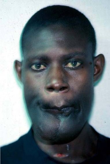
GIANT AMELOBLASTOMA


GIANT AMELOBLASTOMA (CASE PICTURES )